Project

High resolution X-ray microtomography (micro-CT) is a novel technique that allows non-invasive imaging of the internal structure of animals and objects. Virtual cross-sections through the object can be reconstructed allowing 3D rendering.
Micro-CT is becoming essential for studying unique preparations due to its non-destructive nature. This method can be applied to very precious materials, as the object remains intact after the study. Interaction of the high-energy photons with the matter cannot create any radiation damage because the doses that are required to retrieve 3D information about the inner structure are far too small. An important feature of X-ray micro-CT is that the quality of the information does not depend on the distance from the surface. Data about the inner central parts are as accurate as data about the surface. Thanks to these characteristics, several research topics already involve micro-CT as a key tool. Besides applications in materials science and biomedical research, micro-CT has a strong potential to be applied for studying objects belonging to the cultural heritage. Previously the technique was used for investigation of corrosion layers in ancient glasses [1]. Recently, teeth and mandibulae of Neanderthalers and other fossilized structures were scanned and 3D models were reconstructed [2,3].
On top of the hardware possibilities, many complimentary software tools were developed to process the obtained data. As major applications for micro-CT in biomedical research were the analysis of bone and calcified tissue, studies of the spongeous architecture of bones resulted in software that can be applied to the analysis of porous stone that can be corroded in historical building materials. This software takes into account the 3D distribution of pores and not a set of 2D cross-sections such as is the case in classical histology. Therefore it becomes possible to calculate more realistic and additional mesh parameters such as connectivity, anisotropy, average pore sizes etc.
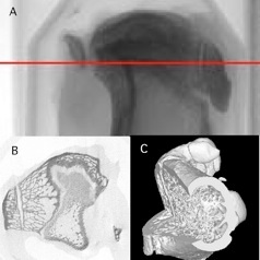
Illustration of high-resolution micro-CT analysis of a bone. A : scout view of the bone. B : virtual sectioning (corresponding to red line on A). C : 3D imaging from stack of virtual cross-sections.
At present, spatial resolution of the method is restricted to 10 microns although it is possible to include nano-CT with submicron resolution to study tiny preparations if necessary.
Normally, contrast between corroded and unaffected layers, and contrast between material and empty pores is sufficient for qualitative analysis of the structures. Therefore, the following prospective directions for micro-CT applications in the domain of cultural heritage are proposed:
-scanning of ancient documents to define porosity and internal damages and correlate them with complimentary studies of the same object;
-fragments of paintings where different layers can be analyzed, the results can be applied for restoration;
-investigations of porosity and the degree of filling in pieces of ancient building materials for preservation studies;
- internal structure and anatomy of fossils and analysis of ancient bones
In all these applications CT can be used as a first non-destructive diagnostic device, providing 3D and spatial information to further extent the analysis with complementary techniques.
To further develop the possibilities of micro-CT, the illuminating spectrum can be altered. Scanning of samples with different excitation wavelength spectrum might help to elucidate the element composition of the samples. Different energy spectra can be provided by variation of X-ray tube anode material. Using of a set of X-ray tubes with different strong characteristic lines allow to estimate the fraction of one element in a mixture of several elements. In this case elemental resolution will be added to spatial resolution. The changes that should be applied to the micro-CT device that will be involved in the project are technically possible and do not require expensive and extensive redesign of the system.
In addition, we are planning to use a wide range of micro-CT instruments to cover various requirements for resolution, object sizes and necessary contrast. The unique chance to unite the power of different scanners will provide us with the possibility to study samples up to 70 mm in diameter with 10 micron resolution and also go down to <1 micron resolution on tiny pieces.
The other effort in 3D imaging will be the development of X-ray fluorescence tomography using synchrotron radiation. This will be described in the WP2 as element distribution is also involved.
References
[1] Roemich H., Lopes E., Mees F., Jacobs P., Cornelis E., van Dijck D., Domenech Carbo T., Micro-computed tomography (mCT) as a new non-destructive tool for the characterisation of archaeological glasses.- In: Proceedings of the 7th International Conference on Non-Destructive Testing and Microanalysis for the Diagnostics and Conservation of the Cultural and Envrionmental Heritage, Antwerp, Belgium / van Grieken R. [edit.], e.a., s.l., , 2002
[2] Semal P., Toussaint M., Maureille B., Rougier H., Crevecoeur I., Balzeau A., Bouchneb L., Louryan S., De Clerck N., Rausin L., Numération des restes humains néandertaliens belges. Préservation patrimoniale et exploitation scientifique, Notae Praehistoricae 25 (2005) 25-38
[3]. Semal P., Cornelissen E., Wannijn L., De Mens van Spy in 3D, Science connection 11 (2006) 35-39
Virtual sectioning and 3D imaging