Project

Elemental analysis
Identification
X-ray fluorescence
P1, P2, P4
X-ray based methods are the most frequently used in the cultural heritage field because they inflict the least damage to the materials/artifacts being examined, as opposed to IR/VIS/UV laser-based or charged-particle based methods of microanalysis, where the rate of local energy deposition is 10-1000 more higher than in the case of X-ray irradiation.
Generally speaking, due to an improvement of performance and miniaturization of X-ray sources, X-ray optics and X-ray fluorescence detectors, a significant improvement in the capabilities of X-ray microprobes has been realized in the last 10 years, with respect to the lowest concentration/mass levels at which meaningful measurements can be performed, the scale/lateral/spatial resolution at which local measurements are possible (line scans, imaging in 2D and 3D) and the quality of the information obtained.
Classical XRF can be done with portable system (WP3) but also in the lab or in synchrotron.
Ion beam techniques
P1, EU1
For several years, the archeologists and art experts call more and more often for experimental techniques in order to obtain quantifiable information allowing to appreciate the antiquity, the environment and the authenticity of the artifacts to be investigated. Among the methods available, the one's using accelerated charged particles take a very important place.
Particle induced X-ray emission (PIXE) is widely used in art and archaeology because of its great advantages: using an external beam, PIXE is easily adaptable to any kind of art object, it provides several information during one experiment, and with a low beam current density, it causes no damage to the target (except in the case of paper-like objects, where a limitation of beam fluence is needed). Moreover, a high sensitivity can be reached.
In the case of paintings, information concerning the elemental concentration of the pigments is a source of precious data. Unfortunately a painted surface is very often composed by several layers of different compositions. The main problem with this kind of samples is the heterogeneity of the interface between the successive layers. Therefore one should be fully aware that the results obtained by any method have to be considered with care. We will use here a specific PIXE atmospheric set-up [1] allowing the separation of the information coming from the different painting layers and presence of degradation. Furthermore, even objects of large size can be investigated with the atmospheric PIGE–PIXE set-up.
Elemental distribution
Quantitative three-dimensional XRF spectroscopy on the microscopic and submicroscopic scale: method development and applications in archaeology
In conventional scanning micro-XRF experiments, an X-ray micro beam is moved in a XY-fashion over the surface of a sample to be examined while X-ray fluorescence spectra are recorded at every point [2]. Since the detection limit of micro-XRF in laboratory equipment is situated at the 10-50 µg/g level while at synchrotron beamlines, sub-ppm MDL-values can be achieved, this type of 2D scanning allows for visualization of major to trace element distributions in various materials. Such maps can be useful, e.g., for studying migration of (trace) constituents during corrosion of historic artifacts etc. [3].
However, considering that energetic X-ray beams (typically 25-30 keV) in some materials can penetrate several hundreds of microns, it is possible also to examine the distribution of elements within artifacts in planes below the surface (in so-called virtual cross-sections) by employing either:
(a) confocal micro-XRF (and its extentions)
i.e. a newly established 3D variant of confocal 2D micro-XRF [4,5], which recently has proven its high potential for the completely non-destructive elemental depth profiling of cultural heritage materials such as paint layer stratigraphies in easel painting [6], enameled tiles, illuminated prints, corroded metallic objects and heavy metals in heterogeneous sediments [7] etc. This method, based on the use of focusing X-ray optics both in the path of the primary beam as well as in the path of the secondary fluorescent radiation traveling between sample and detector, allows to visualize with a spatial resolution of > 10 µm the distribution of elemental species in arbitrarily defined planes within the material/artifact, provided the location of these planes below the object surface is within the sampling depth of the X-ray signals employed.
(b) X-ray fluorescence computer tomography (XFCT),
which involves the coordinated translation and rotation of small samples through (sub)microscopic X-ray beams while X-ray fluorescence signals are being collected and allows to obtained high resolution (typically 100-500 nm) distribution maps in the virtual plane of rotation/translation which is located within the sample under examination [8]. Several tomographic maps may be combined to yield 3D distributions of specific constituents of the samples. Successful XFCT on geological samples (natural diamond-inclusions) has been performed recently by the ESRF ID18F beam line by Partners P2 and P4 providing unique fully three-dimensional information on the submicroscopic distribution of metals in inclusions buried in diamonds [9] and in zeolite crystals [10].
The aim of this work-package is the further development of these techniques towards a fully three-dimensional, quantitative analytical method with lateral resolution levels down to the nanoscopic scales (~50-100 nm) [11,12,13]. The methodological developments described below will be employed to perform non-destructive 3D elemental analysis of unique samples of archaeological or artistic nature of various origin.
This work-package of the present IAP project for the period of 2007 - 2012 is associated with several ongoing ESRF/HASYLAB long-term projects and can be divided in 2 distinct parts:
A. Extension and improvement of confocal micro-XRF towards confocal micro-XAS; extension towards confocal micro-PIXE
Confocal X-ray absorption microspectroscopy. By performing confocal micro-XRF measurements at slightly different energies of the primary micro beam, situated close to the absorption edge of an element of interest, if becomes possible to visualize the distribution of elements in a specific oxidation state (e.g., metallic Cu as in uncorroded Cu-alloys, Cu+ as is cuprite, Cu2+ as in Cu-salts) in three-dimensions. This was recently demonstrated in various contexts by partner P2 (see [7] for the case of As in sediments). Alternatively, chemical state 3D mapping can also be realized by doing repeated XFCT experiments at different energies. Since highly monochromatic X-ray beams are required to do this, such type of measurements are only possible in optimal conditions, i.e., by using state-of-the-art synchrotron undulator sources, highly efficient focusing optics and very large solid-angle X-ray detectors (for XFTC). Thus, in order to implement a facility that will permit these type of measurements to be performed in a routine fashion to cultural heritage problems, the currently available experimental setup at the ESRF-ID18F beamline needs to be optimized for this. These activities will be performed between partners P2, P4 and EU1.
Confocal external beam proton-induced X-ray emission. Another way of extending the application potential of 3D analysis is to combine the confocal detection scheme, which involves collimating/focussing X-ray optics between sample and X-ray detector, with primary sample excitation by a proton micro beam. Such activity will be executed in close collaboration between partners P2 and EU1, with help of partner P1. An efficiently operating confocal micro-PIXE setup, situated in the EU1 laboratory immediately adjacent to one of Europe major musea (the Louvre) would be very useful for preliminary collection of non-destructive depth-profiles of paint layer stratigraphies directly on large panel or easel paintings, allowing ''multiple point'' screening of paint-layer build-up in these paintings prior to any sampling necessary for XFTC or equivalent analyses.
B. Development of sub-micron scanning X-ray fluorescence tomography using refractive optics
Similarly to other scanning micro-beam techniques, the lateral resolution of XFCT is ultimately limited by the employed beam size. Currently, the highest practical lateral resolution level achieved with this technique is in the 0.4 micrometer range with absolute detection limits down to about 10 attogram for the most efficiently excited transition metals [12].
With the ongoing upgrade of the Microfocus beamline (ID13) at the ESRF, beam sizes in the range of 50-100 nm are envisaged for scanning XRD/XRF applications. At this instrument,
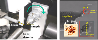
Sub-micron XRF-tomography at ESRF ID18F: experimental setup (left) and SEM image of the investigated zeolite microparticle of ca. 15 m in size (right). Also shown is the reconstructed Cu-map obtained from the central (virtual) cross-section of the particle at a resolution level of 400 nm (right panel).
beam sizes of 69x33 nm have been already demonstrated using two-dimensional X-ray wave-guides [6]. A possible alternative for nanofocusing is based on a special design of “nanofocusing compound refractive lenses” (nanofocusing-CRL’s) produced by the Technical University of Aachen, Germany [11]. These nanofocusing CRLs were the editor’s choice in Science 299, 1628 (2003). Our aim is to reach an up-to-now undemonstrated resolution level below 100x100 nm2, and to demonstrate the feasibility of XRF-tomography at this resolution level in collaboration with Dr. C. Schroer, (Univ. Dresden, Germany). The current generation of nanofocusing CRL’s have recently reached beam sizes down to 47 nm (V) x 55 nm (H) [13]. 3D visualization methods with nanoscopic resolution are showing very high potential for studying the fundamental mechanisms that initiate for instance corrosion processes at (inter)metallic surfaces and interfaces.
More specifically, this phase of the work will also be concerned with the generalisation of accurate analysis from the point and 1D/2D level to quantitative 3D analysis on the basis of X-ray fluorescence computed tomography (XFCT) and its combination with confocal XRF. Feasibility studies and applications of local X-ray fluorescence tomography will be demonstrated, which is achieved by the combination of confocal detection with high-resolution fluorescence tomography. This part of the work is planned at various ESRF beam lines in collaboration with ESRF staff and Dr. C. Schröer (Univ. Dresden). ESRF beamlines ID13, ID18F and ID22 are currently one of the few instruments in the world where sufficiently intense sub-micron hard X-ray beams are available and are equipped for combined sub-micron XRF/XRD analysis [10,12].
An important aspect of XFCT is the tomographic reconstruction based on the accurate modelling of the photon-matter interactions involved. This becomes more important as the spatial resolution of the technique is improved, in particular in view of secondary effects, such as characteristic X-ray emission induced by energetic photoelectrons. This can pose a fundamental limitation to the achievable lateral resolution in case of sub-micron XRF imaging as the energetic photoelectrons from ionisation of lower-Z elements can travel significant distances in the matrix. These phenomena will be studied throroughly by detailed Monte Carlo simulations which are used for tomographic reconstruction. These effects have so far not been included in XRF-tomographic reconstruction techniques.
Up to now, only semi-quantitative XFCT reconstruction algorithms have been demonstrated in the literature, including basic correction for self-absorption effects in the matrix material. These methods need additional calibration procedures for the calculation of the distribution of the absolute concentrations of the sample elements.
In the framework of this work-package the potential of three-dimensional elemental analysis by XFCT will be combined with the quantitative character of the reverse Monte Carlo method [14] to develop a new, ‘no-compromise’ reconstruction for XFCT. The further development of our XRF-simulation model will include the full implementation of new interaction types, namely photoelectron impact ionisation/bremsstrahlung and X-ray resonant Raman scattering (XRRS), which are important for determining the detected sample response in case of monochromatic SR-excitation, improving the accuracy of quantitative XRF-analysis.
This development will provide the possibility of non-destructive quantitative 2D/3D elemental imaging down to submicron scales for delicate and unique samples of archaeological origin.
C. Application and demonstration of the newly developed analysis procedures in the cultural heritage field
Applications of micro-XFCT and confocal XRF in archaeology where the in-situ, non-destructive, contamination free, accurate elemental/structural analysis and the 3D characterization of minor/trace element distributions is important for the full understanding of the origin/source, history, manufacturing details and potential ways of preservation/restauration of the examined materials.
It is important to point out in this respect that the application of such state-of-the-art 3D characterization methods always is taking place in combination with the use of other 2D or 3D characterization methods providing less rich or less detailed information. For example, prior to using either high-resolution confocal-XRF (P2), confocal-XAS (P2) or XFCT (P4), almost invariably, laboratory-based X-ray absorption microtomography or confocal-PIXE (EU1) measurements are likely to be undertaken to explore the structure, internal density variations and compositional heterogeneity of the artifacts/samples to be studied.
Applied studies likely to be undertaken with confocal micro-XRF, micro-XAS and confocal PIXE include the systematic investigation of paint layer stratigraphy and pigment use variations between 15 C 'Flemish primitive' painters such as J. Van Eyck, H. Memling, R. Van der Weyden and others (in combination with screening methods such as portable XRF, mobile Raman spectroscopy, …), mapping of redox gradients in corroding metal alloys (Cu, Fe, …) and in corroding paper documents prepared with ferro-gallic ink while a typical challenging XFCT application will be the submicroscopic investigation of alteration layers at the surface of small pigment grains (smalt, cinnabar, verdigris, …) as a result of contact with (acidic) binding media, causing colour changes.
Ion beam analysis
The development of X-ray lenses used in 3D µ-XRF lead us to investigate the possibility to apply this concept to the PIXE method. This will be developed and tested in collaboration with P2 and the EU1 (see above).
RBS (Rutherford Backscattering Spectrometry) is one of the Ion beam Analysis (IBA) method dedicated to the analysis of multi layers samples and can be, to our mind, easily adapted to the analysis of painted layers [15]. As the other IBA methods in external beam mode, RBS is non destructive, the thickness analysed can be adjusted to the type of sample by changing the nature and the energy of the particle beam (from 3-4 µm for a 3 MeV 4He2+ beam to 50µm for a 5 MeV H+ beam). In a common use RBS is more often use for the analysis of relatively heavy matrices (metal) but it can be very interesting to use it in the domain of painting.
The first point is to evaluate in classical painting the thickness of the coloured paint layer. The use of lead white as preparation layer will be here very useful because of the importance of the lead signal in RBS spectrum, which will be used to calculate the energy loss due to the coloured layer.
In a second point this method will be also available in the evaluation of the thickness of the varnish layer using a particularity of the method when the analysis is made with a proton beam. In this case the signal of light elements especially carbon which usually is very low, is enhanced by nuclear interactions and becomes significant for analysis.
These two points will be very useful to control the quality of the results obtained by the PIXE analysis which can be performed simultaneously with the same beam.
In an other point of view the RBS spectrum can be used to evaluate the homogeneity of a layer and can be a very good mean to detect and understand a degradation process in particular in case of glass, glaze and enamels.
Finally RBS is almost the only non-destructive technique that permits to study the nature and the interactions at the interfaces between different layers. This property will be useful on all types of materials to infer both the material’s nature and the artefact’s fabrication technique and characterize the possible degradation of the polychrome layer and their interactions with the support.
Molecular identification
Raman spectroscopy
Although the Raman effect was for the first time described in 1928, for many years the application of this method was limited to highly specialized applications, the approach was complex and time-consuming and the sensitivity of the technique was low. However, during the history of this technique several instrumental improvements, such as the introduction of new light sources, better detectors and optical elements, made Raman spectroscopy available for new research fields, including art analysis.
Indeed, Raman spectroscopy is a very useful technique for the non-invasive investigation of objects of art [16-19]. Especially its non-destructive character of this molecular spectroscopic technique and its ability to obtain information from micrometer-sized particles are well-appreciated. By using this technique, it is possible to identify mineral pigments as well as organic materials, which illustrates the versatility of the approach: Raman spectroscopy can be used for the investigation of ancient as well as modern art objects.
The introduction of fibre optics in Raman spectroscopy made direct analysis possible, instead of sampling the artefact. Further miniaturization of the instrumentation enabled us to develop mobile equipment for in situ investigation of artefacts. The main advantages of this approach are obvious: no sample has to be taken and the object of art has not to be transported. This makes that the work of art can be examined in a non-destructive way.
In this project, the Raman research group (P3) can contribute in several work packages. The research group’s scientific interests in the field of art analysis are quantitative Raman analysis, the identification of inorganic as well as organic products and the use of combined method approaches. This research is performed on dummies, as well as on artefacts – depending on the research questions on hand.
Raman spectroscopy enables us to obtain the molecular composition of a sample or an area of an artwork. Theoretically, the obtained Raman signal is proportional to the concentration of a (molecular) compound in the sample volume. However, in practice, this approach is not straightforward, since several interferences may interfere: sample inhomogeneity, instrumental instability (laser, optics, detector), the definition of the sample volume, etc. The aim of this project is to find answers to these problems, in order to be able to determine the composition of paint layers in a (semi) quantitative way (WP4). Research will be performed on dummies and the results will be compared with those of other techniques available from partners in the project.
One of the advantageous properties of Raman spectroscopy is its ability to obtain information on the inorganic as well as on the organic compounds in the artefact. However, the identification of the products is based on the comparison with spectra from a reference spectral library and partially on spectral interpretation. Most inorganic compounds are present in the established reference databases, but the reference library needs to be extended for many organic paint compounds (WP4). Spectral interpretation will allow us to study degradation phenomena. This is of high importance to answer specific questions from restorers, since many conservation questions are related to the binding media and varnishes (WP5).
FT-IR
To fully understand the painting technique the identification of the organic compounds, mainly binding media and dyes or lake pigments, is essential. With the exception of some dyes or lake pigments, and in a rare case some binding media that can be identified by Raman spectroscopy or Fourier-transform infrared spectroscopy (FT-IR), all techniques currently used for organic analysis are micro-destructive, in the sense that the sample is consumed during analysis. Gas chromatography-mass spectrometry (GC-MS) delivers detailed information on the compounds present, with an excellent sensitivity so that sampling stays limited. Samples that cannot easily be dissolved can be introduced to the GC by using pyrolysis (Py-GC-MS) as a way of sample introduction. This is not a routine method, due to the sometimes complex and difficult to interpret pyrograms that are registered. Dyes and lakes are also analysed using micro-destructive methods, often high performance liquid chromatography (HPLC) coupled to sensitive detectors like diode array detectors (DAD) or mass spectrometers (MS).
Recently there are also some developments that could allow the determination of organic compounds like binding media or lakes directly on cross-sections. This would be a great step forward since now sampling for binding media or lake analyses is usually carried out by scraping. A major disadvantage is loss of information concerning the spatial distribution of organic compounds in the multi-layered paint system. Also in some cases layers can be so thin that they cannot be separately sampled and results become difficult to interpret. Direct measurement on a cross-section would avoid such problems and can lead to an image of the distribution of (in)organic compounds. One such a method could be precision infrared chemical imaging using a new generation of FT-IR instruments. The technique reveals detailed information on the organic functional group distribution in individual layers of embedded paint cross sections and is used complimentary to visual microscopy and SEM-EDX [20]. The method will be further optimised and applied on some of the many cross-sections available at KIK/IRPA. Also for the study of transformation and degradation phenomena (WP5) this technique can be helpful.
SIMS
Also static secondary ion mass spectrometry (sSIMS) has recently been introduced as an analytical technique for the examination of paint cross sections to obtain simultaneous information about the nature and distribution of pigments and the binding medium from a single sample. sSIMS allows de characterization of the components of the last mono-molecular layer of an organic or inorganic sample in the solid state [21]. The technique is based on the analysis of secondary ions ejected from the surface after bombardment with a beam of primary ions with high energy (in the order of keV). The ion beam can be focussed to sub-micrometer values, making this a micro-analytical method. The static mode, in contradiction to dynamic SIMS, uses a primary ion rate not exceeding 1012 ions cm-2. The value of the method for chemical imaging and identification of oil binding media has recently been shown [22]. In the current research project this method will be adapted and used for visualizing the distribution of proteinacious binding media in cross-sections. Current techniques of identification, support largely on complete hydrolysation of the proteins into amino acids, which are measured with either HPLC or GC-MS after derivatisation. A lot of information is lost, for instance about the structure, matrix effects and degradation processes. The method will also be applied for the analysis of lake pigments directly on cross-sections. At the same time also information concerning oily binding media and pigments present will be obtained. Finally this method will be useful for the study of degradation issues (WP5). All the work with SIMS will be carried out in co-operation with Prof. Patrick Bertrand, specialised in this technique (Unité de physico-chimie et de physique des matériaux, PCPM, Université Catholique de Louvain UCL, betrand@pcpm.ucl.ac.be) [23]
Structural analysis
The history and the properties of materials are deduced not only from their elemental and molecular signatures, but also from their exact mineral phase compositions, and from the structures and the defects of their constituents. X-ray diffraction is an important tool for this research.
The X-ray diffraction laboratory at the C2RMF is equipped with Bruker D5000 X-ray diffractometer with a Gobel mirror. Since two years, a second diffractometer has been developed to characterize paintings and samples at a lower scale: a Rigaku MICROMAX 002 X-ray tube is used with an imaging plate detector. Analyses could be performed with beam sizes of 200µm, 100µm, 30µm.
A specific sample holder has been developed to characterize area on paintings or small cross-sections of painting sample. This system was devised so as to be especially suitable in the Cultural Heritage field, where often artefacts can have large dimensions and curved or rough
surfaces. Therefore, X-ray diffraction has to be performed in reflection and quasi-parallel beam geometry.
Other experiments has been developed with synchrotron radiation at the European Synchrotron Radiation facility (Grenoble). The project is to study the textures and graininess of pigments. We think that the physical properties of pigment grains are related to the artist’s know-how. We have implemented a non-destructive synchrotron X-ray based method, which combines both the quantitative structural content of diffraction and the imaging mode. As a demonstration case, the pigments of a Roman wall painting have been examined [24]. The joined elemental and mineral maps mimic the major features of the painting. Different structural phases made of common atomic elements are differentiated.
References
[1] Weber G., Strivay D., Martinot L., Garnir H.P., Use of PIXE-PIGE under variable incident angle for ancient glass corrosion measurements, Nucl. Instr. & Methods B189 (2002) 350-357
[2] Janssens K.., Adams F., Rindby A., (Eds.), "Microscopic X-ray fluorescence analysis", Chichester : Wiley, 2000. - 419 p. - ISBN 0-471-97426-9
[3] Aerts A., Janssens K., Adams F., Trace level microanalysis of Roman glass from Khirbet Qumran , Israel, Journal of Archaeological science 26 (1999) 883-891
[4] Vincze L., Vekemans B., Brenker F.E., Falkenberg G., Rickers K., Somogyi A.,
Kersten M., Adams F., Three-dimensional trace element analysis by confocal X-ray
microfluorescence imaging, Analytical Chemistry 76 (2004), 6786-6791
[5] Janssens K., Proost K., Falkenberg G., Confocal microscopic X-ray fluorescence at the HASYLAB microfocus beamline: characteristics and possibilities, Spectrochimica Acta B 59 (2004) 1637-1645
[6] Smit Z., Janssens K., Proost K., Langus I., Confocal micro-XRF of paint layers, Nucl. Instr. & Methods B219-220 (2004) 35-40
[7] Denecke M.A., Janssens K., Proost K., Rothe J., Noseck U., Confocal micro-XRF and micro-XAFS studies of uranium speciation in a tertiary sediment from a waste disposal natural analogue site, Env. Sci. Technol. 39 (2005) 2049-2058
[8] Vincze L. et al., X-ray fluorescence microtomography and polycapillary based confocal imaging using synchrotron radiation, “Developments in X-ray tomography IV”, U. Bonse, Ed.; SPIE-Bellingham, Washington, 220-231, (2004)
[9] Brenker F.E., Vincze L., Vekemans B., Nasdala L ., Stachel T., Vollmer C., Kersten M., Somogyi A., Adams F., Joswig W., Harris J.W., Detection of a Ca-rich lithology in the Earth’s deep (> 300 km) convecting mantle, Earth and Planetary Science Letters 236 (2005) 579-587
[10] Terzano R., Spagnuolo M., Medici L., Vekemans B., Vincze L., Janssens K., Ruggiero P., Copper stabilization by zeolite synthesis in polluted soils treated with coal fly ash, Env. Sci. & Technol. 39 (2005) 6280-6287
[11] Pfeiffer F., David C., Burghammer M., Riekel C., Salditt T., Two-Dimensional X-ray Waveguides and Point Sources, Science 297 (2002) 230-234
[12] Vincze L., Nasmov V., Vekemans B., Somogyi A., Drakopoulos M., Adams F., Janssens K., Submicron X-ray fluorescence tomography using polymer compound refractive lenses, J. Anal. At. Spectrometry, submitted (2006)
[13] Schroer C.G. et al., Hard x-ray nanoprobe based on refractive x-ray lenses, Applied Physics Letters 87 (2005) 124103.
[14] Vincze L., Janssens K., Vekemans B., Adams, F., Monte Carlo simulation for X-ray fluorescence spectroscopy (chapter in a book), In: X-Ray Spectrometry Based on Recent Technological Advances, Ed. Kouichi Tsuji, Jasna Injuk and R. Van Grieken, [edit.], Publisher: John Wiley & Sons, Ltd., 435-462, 2004
[15] Mathis F., Moignard B., Pichon L., Dubreuil O., Salomon J., Coupled PIXE and RBS using a 6 MeV He external beam: a new experimental device for particle detection and beam monitoring, Nucl. Instr. & Methods B240 (2005) 532-538
[16] Vandenabeele P., Moens L., Micro-Raman spectroscopy of natural and synthetic indigo samples, The Analyst 128 (2003) 187-193
[17] Vandenabeele P., Raman spectroscopy in art and archaeology, J. Raman Spectroscopy 35 (2004) 607-609
[18] Castro K., Vandenabeele P., Rodriguez-Laso M.D., Moens L., Madariaga J.M., Micro-Raman analysis of coloured lithographs, Anal. Bioanal. Chem. 379 (2004) 674-683
[19] Vandenabeele P., Bode S., Alonso A., Moens L., Raman spectroscopic analysis of the Maya wall paintings in Ek’Balam, Mexico, Spectrochimica Acta A 10 (2005) 2349-2356
[20] van der Weerd J., Microscopic analysis of traditional oil paint, Academisch proefschrift, AMOLF/FOM 2002
[21] Van Vaeck L., Adriaens A., Gijbels R., Static Secondary Ion Mass Spectrometry (S-SIMS), Mass Spectrometry Reviews 18 (1999) 1-47
[22] Keune K., Binding medium, pigments and metal soaps characterised and localised in paint cross-section, Academisch proefschrift, AMOLF/FOM, 2005
[23] Bertrand P., Weng L-T., Time-of-Flight Secondary Ion Mass Spectrometry (ToF-SIMS), Microchimica acta (1996) 13, 167 – 182
[24] Dooryhée E., Anne M., Bardiès I., Hodeau J.-L., Martinetto P., Rondot S., Salomon J., Vaughan G.B.M., Walter P., Non-destructive synchrotron X-ray diffraction mapping of a Roman painting, Applied Physics A 81 (2005) 663-667
Elemental, molecular and structural analysis